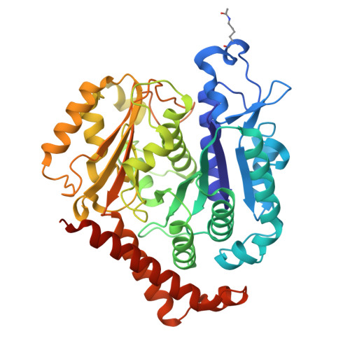
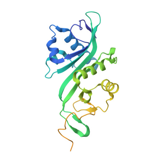
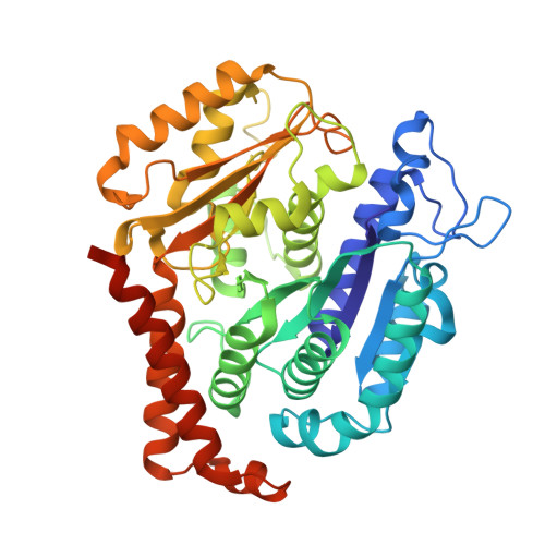
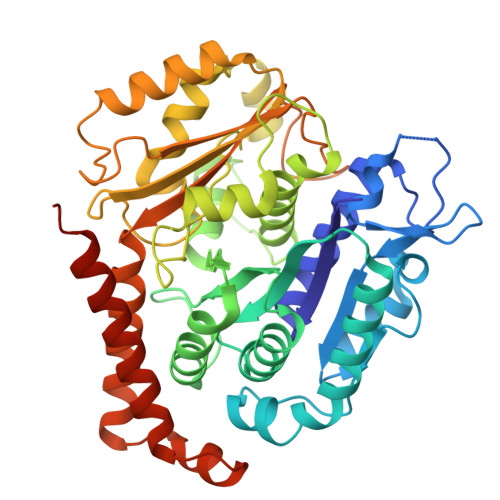
8Y9F

ATAT-2 bound MEC-12/MEC-7 microtubule


Experimental Data Snapshot

  • Method: ELECTRON MICROSCOPY
  • Resolution: 3.30 Å
  • Aggregation State: HELICAL ARRAY 
  • Reconstruction Method: SINGLE PARTICLE 

wwPDB Validation   3D Report Full Report


This is version 1.2 of the entry. See complete history


Literature

Tubulin acetyltransferases access and modify the microtubule luminal K40 residue through anchors in taxane-binding pockets.

Luo, J.Lam, W.H.Yu, D.Chao, V.C.Zopfi, M.N.Khoo, C.J.Zhao, C.Yan, S.Liu, Z.Li, X.D.Zheng, C.Zhai, Y.Ti, S.C.

(2024) Nat Struct Mol Biol 

  • DOI: https://doi.org/10.1038/s41594-024-01406-3
  • Primary Citation of Related Structures:  
    8Y9F, 8YAJ, 8YAL, 8YAR

  • PubMed Abstract: 

    Acetylation at α-tubulin K40 is the sole post-translational modification preferred to occur inside the lumen of hollow cylindrical microtubules. However, how tubulin acetyltransferases access the luminal K40 in micrometer-long microtubules remains unknown. Here, we use cryo-electron microscopy and single-molecule reconstitution assays to reveal the enzymatic mechanism for tubulin acetyltransferases to modify K40 in the lumen. One tubulin acetyltransferase spans across the luminal lattice, with the catalytic core docking onto two α-tubulins and the enzyme's C-terminal domain occupying the taxane-binding pockets of two β-tubulins. The luminal accessibility and enzyme processivity of tubulin acetyltransferases are inhibited by paclitaxel, a microtubule-stabilizing chemotherapeutic agent. Characterizations using recombinant tubulins mimicking preacetylated and postacetylated K40 show the crosstalk between microtubule acetylation states and the cofactor acetyl-CoA in enzyme turnover. Our findings provide crucial insights into the conserved multivalent interactions involving α- and β-tubulins to acetylate the confined microtubule lumen.


  • Organizational Affiliation

    School of Biomedical Sciences, Faculty of Medicine, The University of Hong Kong, Hong Kong SAR, China.


Macromolecules
Find similar proteins by:  (by identity cutoff)  |  3D Structure
Entity ID: 1
MoleculeChains Sequence LengthOrganismDetailsImage
Tubulin alpha-3 chainA [auth B]450Caenorhabditis elegansMutation(s): 0 
Gene Names: mec-12tba-3C44B11.3
EC: 3.6.5
UniProt
Find proteins for P91910 (Caenorhabditis elegans)
Explore P91910 
Go to UniProtKB:  P91910
Entity Groups  
Sequence Clusters30% Identity50% Identity70% Identity90% Identity95% Identity100% Identity
UniProt GroupP91910
Sequence Annotations
Expand
  • Reference Sequence
Find similar proteins by:  (by identity cutoff)  |  3D Structure
Entity ID: 2
MoleculeChains Sequence LengthOrganismDetailsImage
Alpha-tubulin N-acetyltransferase 2B [auth C],
F [auth I]
263Caenorhabditis elegansMutation(s): 0 
Gene Names: atat-2W06B11.1
EC: 2.3.1.108
UniProt
Find proteins for Q23192 (Caenorhabditis elegans)
Explore Q23192 
Go to UniProtKB:  Q23192
Entity Groups  
Sequence Clusters30% Identity50% Identity70% Identity90% Identity95% Identity100% Identity
UniProt GroupQ23192
Sequence Annotations
Expand
  • Reference Sequence
Find similar proteins by:  (by identity cutoff)  |  3D Structure
Entity ID: 3
MoleculeChains Sequence LengthOrganismDetailsImage
Tubulin beta-1 chainC [auth D],
E [auth F]
441Caenorhabditis elegansMutation(s): 0 
Gene Names: mec-7ZK154.3
UniProt
Find proteins for P12456 (Caenorhabditis elegans)
Explore P12456 
Go to UniProtKB:  P12456
Entity Groups  
Sequence Clusters30% Identity50% Identity70% Identity90% Identity95% Identity100% Identity
UniProt GroupP12456
Sequence Annotations
Expand
  • Reference Sequence
Find similar proteins by:  (by identity cutoff)  |  3D Structure
Entity ID: 4
MoleculeChains Sequence LengthOrganismDetailsImage
Tubulin alpha-3 chainD [auth E]450Caenorhabditis elegansMutation(s): 0 
Gene Names: mec-12tba-3C44B11.3
EC: 3.6.5
UniProt
Find proteins for P91910 (Caenorhabditis elegans)
Explore P91910 
Go to UniProtKB:  P91910
Entity Groups  
Sequence Clusters30% Identity50% Identity70% Identity90% Identity95% Identity100% Identity
UniProt GroupP91910
Sequence Annotations
Expand
  • Reference Sequence
Experimental Data & Validation

Experimental Data

  • Method: ELECTRON MICROSCOPY
  • Resolution: 3.30 Å
  • Aggregation State: HELICAL ARRAY 
  • Reconstruction Method: SINGLE PARTICLE 
EM Software:
TaskSoftware PackageVersion
MODEL REFINEMENTPHENIX1.19-4092
RECONSTRUCTIONcryoSPARC4.1.2

Structure Validation

View Full Validation Report


Entry History & Funding Information

Deposition Data


Funding OrganizationLocationGrant Number
Not funded--

Revision History  (Full details and data files)

  • Version 1.0: 2024-11-06
    Type: Initial release
  • Version 1.1: 2024-11-13
    Changes: Data collection, Database references
  • Version 1.2: 2024-11-27
    Changes: Data collection