8B99

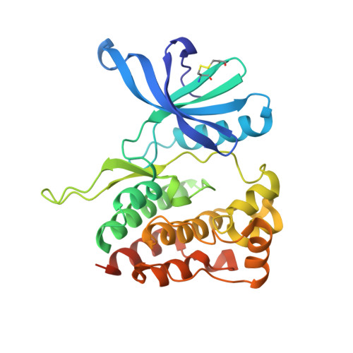
Crystal structure of JAK2 JH2-V617F in complex with JNJ-7706621


Experimental Data Snapshot

  • Method: X-RAY DIFFRACTION
  • Resolution: 1.60 Å
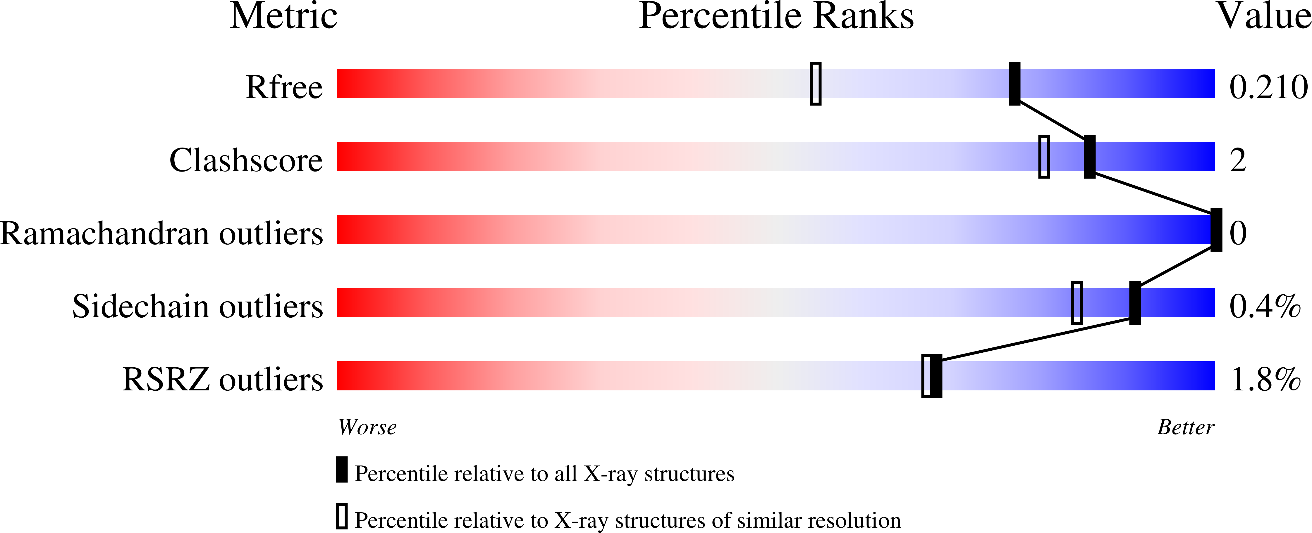
  • R-Value Free: 0.211 
  • R-Value Work: 0.189 
  • R-Value Observed: 0.190 

Starting Model: experimental
View more details

wwPDB Validation   3D Report Full Report


Ligand Structure Quality Assessment 


This is version 1.2 of the entry. See complete history


Literature

Identification of Novel Small Molecule Ligands for JAK2 Pseudokinase Domain.

Virtanen, A.T.Haikarainen, T.Sampathkumar, P.Palmroth, M.Liukkonen, S.Liu, J.Nekhotiaeva, N.Hubbard, S.R.Silvennoinen, O.

(2023) Pharmaceuticals (Basel) 16

  • DOI: https://doi.org/10.3390/ph16010075
  • Primary Citation of Related Structures:  
    8B8N, 8B8U, 8B99, 8B9E, 8B9H, 8BA2, 8BA3, 8BA4, 8BAB, 8BAK, 8EX0, 8EX1, 8EX2

  • PubMed Abstract: 

    Hyperactive mutation V617F in the JAK2 regulatory pseudokinase domain (JH2) is prevalent in patients with myeloproliferative neoplasms. Here, we identified novel small molecules that target JH2 of JAK2 V617F and characterized binding via biochemical and structural approaches. Screening of 107,600 small molecules resulted in identification of 55 binders to the ATP-binding pocket of recombinant JAK2 JH2 V617F protein at a low hit rate of 0.05%, which indicates unique structural characteristics of the JAK2 JH2 ATP-binding pocket. Selected hits and structural analogs were further assessed for binding to JH2 and JH1 (kinase) domains of JAK family members (JAK1-3, TYK2) and for effects on MPN model cell viability. Crystal structures were determined with JAK2 JH2 wild-type and V617F. The JH2-selective binders were identified in diaminotriazole, diaminotriazine, and phenylpyrazolo-pyrimidone chemical entities, but they showed low-affinity, and no inhibition of MPN cells was detected, while compounds binding to both JAK2 JH1 and JH2 domains inhibited MPN cell viability. X-ray crystal structures of protein-ligand complexes indicated generally similar binding modes between the ligands and V617F or wild-type JAK2. Ligands of JAK2 JH2 V617F are applicable as probes in JAK-STAT research, and SAR optimization combined with structural insights may yield higher-affinity inhibitors with biological activity.


  • Organizational Affiliation

    Faculty of Medicine and Health Technology, Tampere University, 33014 Tampere, Finland.


Macromolecules
Find similar proteins by:  (by identity cutoff)  |  3D Structure
Entity ID: 1
MoleculeChains Sequence LengthOrganismDetailsImage
Tyrosine-protein kinase JAK2289Homo sapiensMutation(s): 3 
Gene Names: JAK2
EC: 2.7.10.2
UniProt & NIH Common Fund Data Resources
Find proteins for O60674 (Homo sapiens)
Explore O60674 
Go to UniProtKB:  O60674
PHAROS:  O60674
GTEx:  ENSG00000096968 
Entity Groups  
Sequence Clusters30% Identity50% Identity70% Identity90% Identity95% Identity100% Identity
UniProt GroupO60674
Sequence Annotations
Expand
  • Reference Sequence
Small Molecules
Binding Affinity Annotations 
IDSourceBinding Affinity
SKE BindingDB:  8B99 Ki: 2.20e+5 (nM) from 1 assay(s)
Kd: min: 31, max: 800 (nM) from 9 assay(s)
IC50: min: 1, max: 2960 (nM) from 2 assay(s)
Experimental Data & Validation

Experimental Data

  • Method: X-RAY DIFFRACTION
  • Resolution: 1.60 Å
  • R-Value Free: 0.211 
  • R-Value Work: 0.189 
  • R-Value Observed: 0.190 
  • Space Group: P 21 21 21
Unit Cell:
Length ( Å )Angle ( ˚ )
a = 53.413α = 90
b = 56.724β = 90
c = 114.509γ = 90
Software Package:
Software NamePurpose
GDAdata collection
PHENIXrefinement
XDSdata reduction
XDSdata scaling
PHASERphasing

Structure Validation

View Full Validation Report


Ligand Structure Quality Assessment 


Entry History & Funding Information

Deposition Data


Funding OrganizationLocationGrant Number
Academy of FinlandFinland--

Revision History  (Full details and data files)

  • Version 1.0: 2023-02-01
    Type: Initial release
  • Version 1.1: 2024-02-07
    Changes: Data collection, Refinement description
  • Version 1.2: 2024-10-16
    Changes: Structure summary