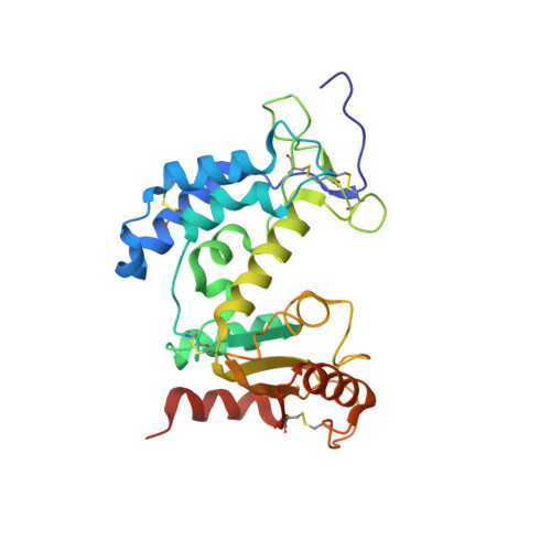
6EDR

Crystal Structure of Human CD38 in Complex with 4'-Thioribose NAD+


Experimental Data Snapshot

  • Method: X-RAY DIFFRACTION
  • Resolution: 2.40 Å
  • R-Value Free: 0.262 
  • R-Value Work: 0.216 
  • R-Value Observed: 0.218 

Starting Model: experimental
View more details

wwPDB Validation   3D Report Full Report


Ligand Structure Quality Assessment 


This is version 2.1 of the entry. See complete history


Literature

Facile chemoenzymatic synthesis of a novel stable mimic of NAD.

Dai, Z.Zhang, X.N.Nasertorabi, F.Cheng, Q.Pei, H.Louie, S.G.Stevens, R.C.Zhang, Y.

(2018) Chem Sci 9: 8337-8342

  • DOI: https://doi.org/10.1039/c8sc03899f
  • Primary Citation of Related Structures:  
    6EDR

  • PubMed Abstract: 

    Nicotinamide adenine dinucleotide (NAD + ) is an essential cofactor participating in a variety of important enzyme-catalyzed physiological and pathophysiological processes. Analogues of NAD + provide key and valuable agents for investigating NAD + -dependent enzymes. In this study, we report the preparation of a novel stable NAD + mimic, 4'-thioribose NAD + (S-NAD + ), using a facile and efficient chemoenzymatic approach. Substrate activity assays indicated the resulting S-NAD + is chemically inert to human CD38 and sirtuin 2 enzymes, but capable of participating in redox reactions in a manner similar to NAD + . X-ray crystallographic analysis revealed binding of S-NAD + to the active site of human CD38 and critical residues involved in leaving group activation and catalysis. By more closely mimicking NAD + in geometry and electrostatics, the generated S-NAD + offers a unique and important tool that can be extended to study enzymes utilizing NAD + .


  • Organizational Affiliation

    Department of Pharmacology and Pharmaceutical Sciences , School of Pharmacy , University of Southern California , 1985 Zonal Ave , Los Angeles , CA 90089 , USA . Email: [email protected].


Macromolecules
Find similar proteins by:  (by identity cutoff)  |  3D Structure
Entity ID: 1
MoleculeChains Sequence LengthOrganismDetailsImage
ADP-ribosyl cyclase/cyclic ADP-ribose hydrolase 1
A, B
257Homo sapiensMutation(s): 4 
Gene Names: CD38
EC: 3.2.2.6 (PDB Primary Data), 2.4.99.20 (PDB Primary Data), 3.2.2 (UniProt)
UniProt & NIH Common Fund Data Resources
Find proteins for P28907 (Homo sapiens)
Explore P28907 
Go to UniProtKB:  P28907
PHAROS:  P28907
GTEx:  ENSG00000004468 
Entity Groups  
Sequence Clusters30% Identity50% Identity70% Identity90% Identity95% Identity100% Identity
UniProt GroupP28907
Sequence Annotations
Expand
  • Reference Sequence
Small Molecules
Ligands 1 Unique
IDChains Name / Formula / InChI Key2D Diagram3D Interactions
ZNA (Subject of Investigation/LOI)
Query on ZNA

Download Ideal Coordinates CCD File 
C [auth A],
D [auth B]
[[(2~{R},3~{S},4~{R},5~{R})-5-(3-aminocarbonylpyridin-1-ium-1-yl)-3,4-bis(oxidanyl)thiolan-2-yl]methoxy-oxidanyl-phosphoryl] [(2~{R},3~{S},4~{R},5~{R})-5-(6-aminopurin-9-yl)-3,4-bis(oxidanyl)oxolan-2-yl]methyl hydrogen phosphate
C21 H28 N7 O13 P2 S
YLEHCOOOOOOSDX-NNYOXOHSSA-O
Experimental Data & Validation

Experimental Data

  • Method: X-RAY DIFFRACTION
  • Resolution: 2.40 Å
  • R-Value Free: 0.262 
  • R-Value Work: 0.216 
  • R-Value Observed: 0.218 
  • Space Group: P 1 21 1
Unit Cell:
Length ( Å )Angle ( ˚ )
a = 57.742α = 90
b = 51.106β = 96.89
c = 100.674γ = 90
Software Package:
Software NamePurpose
REFMACrefinement
XDSdata reduction
SCALAdata scaling
PHASERphasing
PDB_EXTRACTdata extraction

Structure Validation

View Full Validation Report


Ligand Structure Quality Assessment 


Entry History & Funding Information

Deposition Data


Funding OrganizationLocationGrant Number
Other privateUnited StatesV2016-021
National Institutes of Health/National Institute of Diabetes and Digestive and Kidney Disease (NIH/NIDDK)United StatesP30DK048522

Revision History  (Full details and data files)

  • Version 1.0: 2018-11-21
    Type: Initial release
  • Version 1.1: 2019-11-13
    Changes: Database references
  • Version 1.2: 2019-12-25
    Changes: Author supporting evidence
  • Version 1.3: 2023-10-11
    Changes: Data collection, Database references, Refinement description
  • Version 2.0: 2024-05-15
    Changes: Derived calculations, Non-polymer description, Structure summary
  • Version 2.1: 2024-11-20
    Changes: Structure summary