5CMV

Ultrafast dynamics in myoglobin: dark-state, CO-ligated structure


Experimental Data Snapshot

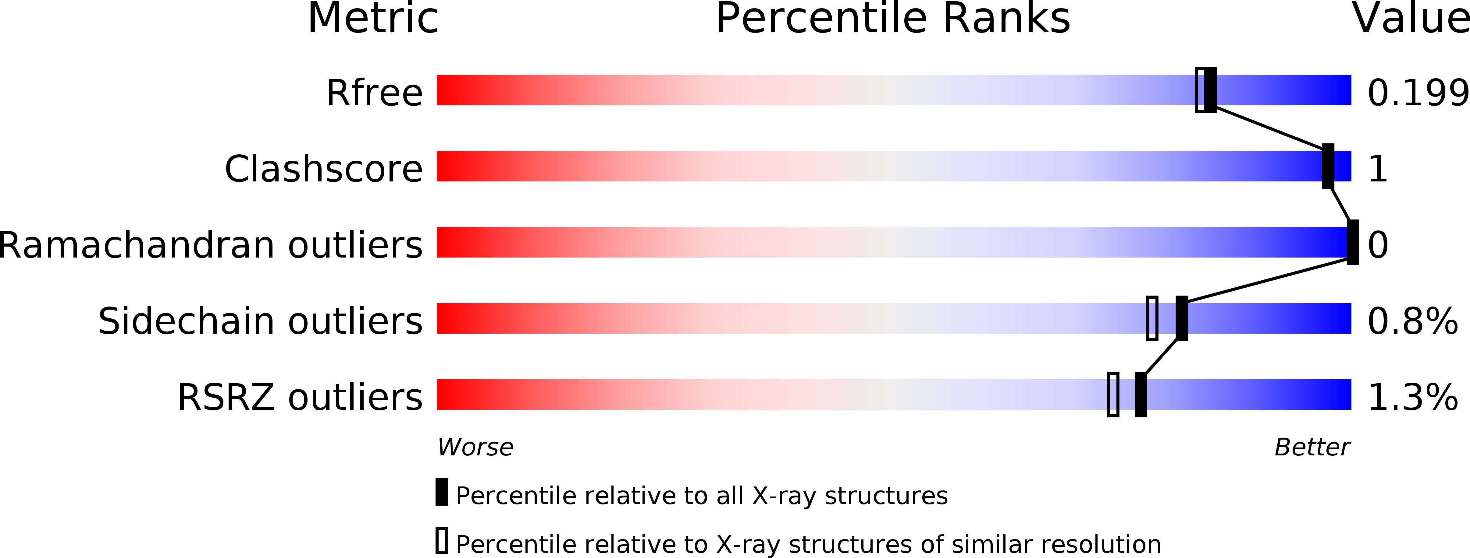
  • Method: X-RAY DIFFRACTION
  • Resolution: 1.80 Å
  • R-Value Free: 0.204 
  • R-Value Work: 0.160 
  • R-Value Observed: 0.162 

Starting Model: experimental
View more details

wwPDB Validation   3D Report Full Report


This is version 1.5 of the entry. See complete history


Literature

Direct observation of ultrafast collective motions in CO myoglobin upon ligand dissociation.

Barends, T.R.Foucar, L.Ardevol, A.Nass, K.Aquila, A.Botha, S.Doak, R.B.Falahati, K.Hartmann, E.Hilpert, M.Heinz, M.Hoffmann, M.C.Kofinger, J.Koglin, J.E.Kovacsova, G.Liang, M.Milathianaki, D.Lemke, H.T.Reinstein, J.Roome, C.M.Shoeman, R.L.Williams, G.J.Burghardt, I.Hummer, G.Boutet, S.Schlichting, I.

(2015) Science 350: 445-450

  • DOI: https://doi.org/10.1126/science.aac5492
  • Primary Citation of Related Structures:  
    5CMV, 5CN4, 5CN5, 5CN6, 5CN7, 5CN8, 5CN9, 5CNB, 5CNC, 5CND, 5CNE, 5CNF, 5CNG, 5D5R

  • PubMed Abstract: 

    The hemoprotein myoglobin is a model system for the study of protein dynamics. We used time-resolved serial femtosecond crystallography at an x-ray free-electron laser to resolve the ultrafast structural changes in the carbonmonoxy myoglobin complex upon photolysis of the Fe-CO bond. Structural changes appear throughout the protein within 500 femtoseconds, with the C, F, and H helices moving away from the heme cofactor and the E and A helices moving toward it. These collective movements are predicted by hybrid quantum mechanics/molecular mechanics simulations. Together with the observed oscillations of residues contacting the heme, our calculations support the prediction that an immediate collective response of the protein occurs upon ligand dissociation, as a result of heme vibrational modes coupling to global modes of the protein.


  • Organizational Affiliation

    Max-Planck-Institut für Medizinische Forschung, Jahnstraße 29, 69120 Heidelberg, Germany. [email protected] [email protected].


Macromolecules
Find similar proteins by:  (by identity cutoff)  |  3D Structure
Entity ID: 1
MoleculeChains Sequence LengthOrganismDetailsImage
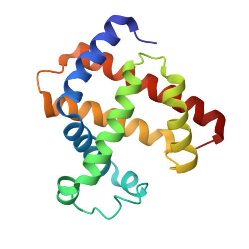
Myoglobin152Equus caballusMutation(s): 0 
EC: 1.11.1 (UniProt), 1.7 (UniProt)
UniProt
Find proteins for P68082 (Equus caballus)
Explore P68082 
Go to UniProtKB:  P68082
Entity Groups  
Sequence Clusters30% Identity50% Identity70% Identity90% Identity95% Identity100% Identity
UniProt GroupP68082
Sequence Annotations
Expand
  • Reference Sequence
Experimental Data & Validation

Experimental Data

  • Method: X-RAY DIFFRACTION
  • Resolution: 1.80 Å
  • R-Value Free: 0.204 
  • R-Value Work: 0.160 
  • R-Value Observed: 0.162 
  • Space Group: P 1 21 1
Unit Cell:
Length ( Å )Angle ( ˚ )
a = 63.6α = 90
b = 28.8β = 106.5
c = 35.6γ = 90
Software Package:
Software NamePurpose
PHENIXrefinement
PHENIXphasing
CrystFELdata reduction
CrystFELdata scaling

Structure Validation

View Full Validation Report


Entry History 

Revision History  (Full details and data files)

  • Version 1.0: 2015-09-16
    Type: Initial release
  • Version 1.1: 2015-09-23
    Changes: Database references
  • Version 1.2: 2015-11-04
    Changes: Database references
  • Version 1.3: 2018-01-24
    Changes: Data collection
  • Version 1.4: 2018-11-14
    Changes: Data collection
  • Version 1.5: 2024-01-10
    Changes: Data collection, Database references, Refinement description