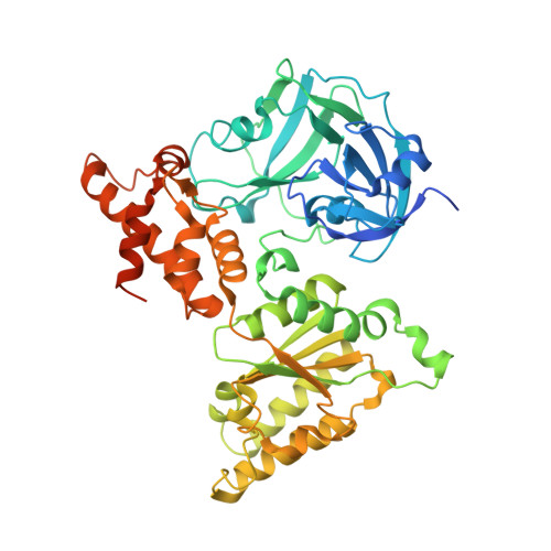
4KOD

Structure of p97 N-D1 R155H mutant in complex with ADP


Experimental Data Snapshot

  • Method: X-RAY DIFFRACTION
  • Resolution: 2.96 Å
  • R-Value Free: 0.291 
  • R-Value Work: 0.268 
  • R-Value Observed: 0.270 

wwPDB Validation   3D Report Full Report


Ligand Structure Quality Assessment 


This is version 1.2 of the entry. See complete history


Literature

Altered Intersubunit Communication Is the Molecular Basis for Functional Defects of Pathogenic p97 Mutants.

Tang, W.K.Xia, D.

(2013) J Biol Chem 288: 36624-36635

  • DOI: https://doi.org/10.1074/jbc.M113.488924
  • Primary Citation of Related Structures:  
    4KLN, 4KO8, 4KOD

  • PubMed Abstract: 

    The human AAA ATPase p97 is a molecular chaperone essential in cellular proteostasis. Single amino acid substitutions in p97 have been linked to a clinical multiple-disorder condition known as inclusion body myopathy associated with Paget's disease of the bone and frontotemporal dementia. How the mutations affect the molecular mechanism that governs the function of p97 remains unclear. Here, we show that within the hexameric ring of a mutant p97, D1 domains fail to regulate their respective nucleotide-binding states, as evidenced by the lower amount of prebound ADP, weaker ADP binding affinity, full occupancy of adenosine-5'-O-(3-thiotriphosphate) binding, and elevated overall ATPase activity, indicating a loss of communication among subunits. Defective communication between subunits is further illustrated by altered conformation in the side chain of residue Phe-360 that probes into the nucleotide-binding pocket from a neighboring subunit. Consequently, conformations of N domains in a hexameric ring of a mutant p97 become uncoordinated, thus impacting its ability to process substrate.


  • Organizational Affiliation

    From the Laboratory of Cell Biology, Center for Cancer Research, NCI, National Institutes of Health, Bethesda, Maryland 20892.


Macromolecules
Find similar proteins by:  (by identity cutoff)  |  3D Structure
Entity ID: 1
MoleculeChains Sequence LengthOrganismDetailsImage
Transitional endoplasmic reticulum ATPase
A, B, C, D, E
A, B, C, D, E, F, G, H, I, J, K, L
489Homo sapiensMutation(s): 1 
Gene Names: p97VCP
EC: 3.6.4.6
UniProt & NIH Common Fund Data Resources
Find proteins for P55072 (Homo sapiens)
Explore P55072 
Go to UniProtKB:  P55072
PHAROS:  P55072
GTEx:  ENSG00000165280 
Entity Groups  
Sequence Clusters30% Identity50% Identity70% Identity90% Identity95% Identity100% Identity
UniProt GroupP55072
Sequence Annotations
Expand
  • Reference Sequence
Small Molecules
Ligands 1 Unique
IDChains Name / Formula / InChI Key2D Diagram3D Interactions
ADP
Query on ADP

Download Ideal Coordinates CCD File 
M [auth A]
N [auth B]
O [auth C]
P [auth D]
Q [auth E]
M [auth A],
N [auth B],
O [auth C],
P [auth D],
Q [auth E],
R [auth F],
S [auth G],
T [auth H],
U [auth I],
V [auth J],
W [auth K],
X [auth L]
ADENOSINE-5'-DIPHOSPHATE
C10 H15 N5 O10 P2
XTWYTFMLZFPYCI-KQYNXXCUSA-N
Binding Affinity Annotations 
IDSourceBinding Affinity
ADP PDBBind:  4KOD Kd: 4370 (nM) from 1 assay(s)
Experimental Data & Validation

Experimental Data

  • Method: X-RAY DIFFRACTION
  • Resolution: 2.96 Å
  • R-Value Free: 0.291 
  • R-Value Work: 0.268 
  • R-Value Observed: 0.270 
  • Space Group: P 21 21 21
Unit Cell:
Length ( Å )Angle ( ˚ )
a = 146.52α = 90
b = 170.737β = 90
c = 256.601γ = 90
Software Package:
Software NamePurpose
HKL-2000data collection
MOLREPphasing
REFMACrefinement
DENZOdata reduction
SCALEPACKdata scaling

Structure Validation

View Full Validation Report


Ligand Structure Quality Assessment 


Entry History 

Deposition Data

Revision History  (Full details and data files)

  • Version 1.0: 2013-11-13
    Type: Initial release
  • Version 1.1: 2014-01-08
    Changes: Database references
  • Version 1.2: 2024-02-28
    Changes: Data collection, Database references, Derived calculations, Refinement description