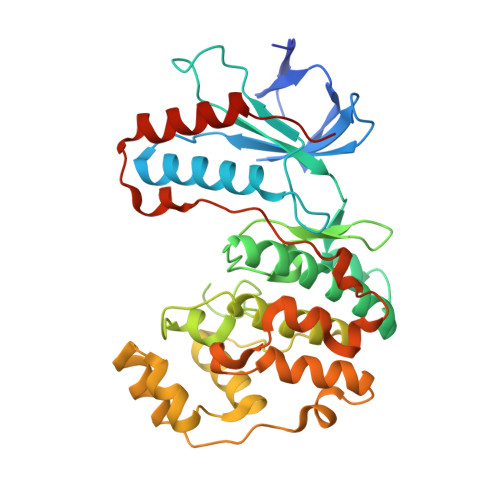
3FC1

Crystal structure of p38 kinase bound to pyrimido-pyridazinone inhibitor


Experimental Data Snapshot

  • Method: X-RAY DIFFRACTION
  • Resolution: 2.40 Å
  • R-Value Free: 0.251 
  • R-Value Work: 0.199 
  • R-Value Observed: 0.204 

wwPDB Validation   3D Report Full Report


Ligand Structure Quality Assessment 


This is version 1.2 of the entry. See complete history


Literature

Evaluating the molecular mechanics poisson-boltzmann surface area free energy method using a congeneric series of ligands to p38 MAP kinase.

Pearlman, D.A.

(2005) J Med Chem 48: 7796-7807

  • DOI: https://doi.org/10.1021/jm050306m
  • Primary Citation of Related Structures:  
    3FC1

  • PubMed Abstract: 

    The recently described molecular mechanics Poisson-Boltzmann surface area (MM-PBSA) method for calculating free energies is applied to a congeneric series of 16 ligands to p38 MAP kinase whose binding constants span approximately 2 orders of magnitude. These compounds have previously been used to test and compare other free energy calculation methods, including thermodynamic integration (TI), OWFEG, ChemScore, PLPScore, and Dock Energy Score. We find that the MM-PBSA performs relatively poorly for this set of ligands, yielding results much inferior to those from TI or OWFEG, inferior to Dock Energy Score, and not appreciably better than ChemScore or PLPScore but at an appreciably larger computational cost than any of these other methods. This suggests that one should be selective in applying the MM-PBSA method and that for systems that are amenable to other free energy approaches, these other approaches may be preferred. We also examine the single simulation approximation for MM-PBSA, whereby the required ligand and protein trajectories are extracted from a single MD simulation rather than two separate MD runs. This assumption, sometimes used to speed the MM-PBSA calculation, is found to yield significantly inferior results with only a moderate net percentage reduction in total simulation time.


  • Organizational Affiliation

    150 Jason Street, Arlington, Massachusetts 02476, USA. [email protected]


Macromolecules
Find similar proteins by:  (by identity cutoff)  |  3D Structure
Entity ID: 1
MoleculeChains Sequence LengthOrganismDetailsImage
Mitogen-activated protein kinase 14A [auth X]366Homo sapiensMutation(s): 0 
Gene Names: MAPK14CSBPCSBP1CSBP2CSPB1MXI2
EC: 2.7.11.24
UniProt & NIH Common Fund Data Resources
Find proteins for Q16539 (Homo sapiens)
Explore Q16539 
Go to UniProtKB:  Q16539
PHAROS:  Q16539
GTEx:  ENSG00000112062 
Entity Groups  
Sequence Clusters30% Identity50% Identity70% Identity90% Identity95% Identity100% Identity
UniProt GroupQ16539
Sequence Annotations
Expand
  • Reference Sequence
Small Molecules
Ligands 2 Unique
IDChains Name / Formula / InChI Key2D Diagram3D Interactions
52P
Query on 52P

Download Ideal Coordinates CCD File 
C [auth X]5-(2,6-dichlorophenyl)-2-[(2,4-difluorophenyl)sulfanyl]-6H-pyrimido[1,6-b]pyridazin-6-one
C19 H9 Cl2 F2 N3 O S
VEPKQEUBKLEPRA-UHFFFAOYSA-N
CL
Query on CL

Download Ideal Coordinates CCD File 
B [auth X]CHLORIDE ION
Cl
VEXZGXHMUGYJMC-UHFFFAOYSA-M
Binding Affinity Annotations 
IDSourceBinding Affinity
52P BindingDB:  3FC1 Ki: 0.8 (nM) from 1 assay(s)
Kd: min: 2.8, max: 1.00e+4 (nM) from 3 assay(s)
IC50: min: 0.8, max: 5300 (nM) from 14 assay(s)
PDBBind:  3FC1 IC50: 10 (nM) from 1 assay(s)
Experimental Data & Validation

Experimental Data

  • Method: X-RAY DIFFRACTION
  • Resolution: 2.40 Å
  • R-Value Free: 0.251 
  • R-Value Work: 0.199 
  • R-Value Observed: 0.204 
  • Space Group: P 21 21 21
Unit Cell:
Length ( Å )Angle ( ˚ )
a = 45.697α = 90
b = 85.958β = 90
c = 123.271γ = 90
Software Package:
Software NamePurpose
CrystalCleardata collection
X-PLORmodel building
REFMACrefinement
DENZOdata reduction
SCALEPACKdata scaling
X-PLORphasing

Structure Validation

View Full Validation Report


Ligand Structure Quality Assessment 


Entry History 

Deposition Data

Revision History  (Full details and data files)

  • Version 1.0: 2008-12-09
    Type: Initial release
  • Version 1.1: 2011-07-13
    Changes: Advisory, Version format compliance
  • Version 1.2: 2023-12-27
    Changes: Data collection, Database references, Derived calculations