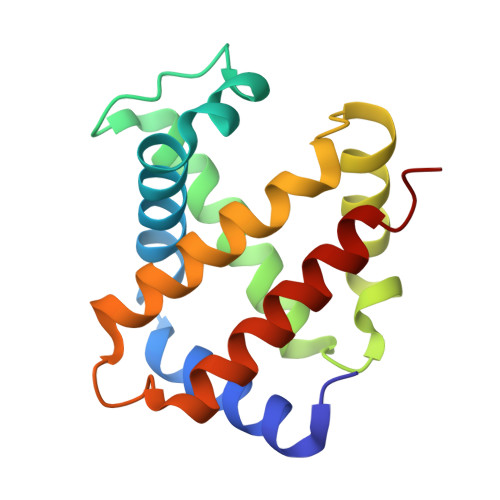
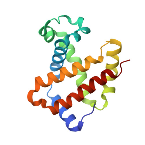
6KAE

Crosslinked alpha(Fe-CO)-beta(Ni) human hemoglobin A in the T quaternary structure at 95 K: Light


Experimental Data Snapshot

  • Method: X-RAY DIFFRACTION
  • Resolution: 1.45 Å
  • R-Value Free: 0.200 
  • R-Value Work: 0.174 
  • R-Value Observed: 0.175 

Starting Model: experimental
View more details

wwPDB Validation   3D Report Full Report


Ligand Structure Quality Assessment 


This is version 1.5 of the entry. See complete history


Literature

Direct observation of ligand migration within human hemoglobin at work.

Shibayama, N.Sato-Tomita, A.Ohki, M.Ichiyanagi, K.Park, S.Y.

(2020) Proc Natl Acad Sci U S A 117: 4741-4748

  • DOI: https://doi.org/10.1073/pnas.1913663117
  • Primary Citation of Related Structures:  
    6KA9, 6KAE, 6KAH, 6KAI, 6KAO, 6KAP, 6KAQ, 6KAR, 6KAS, 6KAT, 6KAU, 6KAV, 6L5V, 6L5W, 6L5X, 6L5Y, 6LCW, 6LCX

  • PubMed Abstract: 

    Hemoglobin is one of the best-characterized proteins with respect to structure and function, but the internal ligand diffusion pathways remain obscure and controversial. Here we captured the CO migration processes in the tense (T), relaxed (R), and second relaxed (R2) quaternary structures of human hemoglobin by crystallography using a high-repetition pulsed laser technique at cryogenic temperatures. We found that in each quaternary structure, the photodissociated CO molecules migrate along distinct pathways in the α and β subunits by hopping between the internal cavities with correlated side chain motions of large nonpolar residues, such as α14Trp(A12), α105Leu(G12), β15Trp(A12), and β71Phe(E15). We also observe electron density evidence for the distal histidine [α58/β63His(E7)] swing-out motion regardless of the quaternary structure, although less evident in α subunits than in β subunits, suggesting that some CO molecules have escaped directly through the E7 gate. Remarkably, in T-state Fe(II)-Ni(II) hybrid hemoglobins in which either the α or β subunits contain Ni(II) heme that cannot bind CO, the photodissociated CO molecules not only dock at the cavities in the original Fe(II) subunit, but also escape from the protein matrix and enter the cavities in the adjacent Ni(II) subunit even at 95 K, demonstrating the high gas permeability and porosity of the hemoglobin molecule. Our results provide a comprehensive picture of ligand movements in hemoglobin and highlight the relevance of cavities, nonpolar residues, and distal histidines in facilitating the ligand migration.


  • Organizational Affiliation

    Division of Biophysics, Department of Physiology, Jichi Medical University, Tochigi, 329-0498 Shimotsuke, Japan; [email protected].


Macromolecules
Find similar proteins by:  (by identity cutoff)  |  3D Structure
Entity ID: 1
MoleculeChains Sequence LengthOrganismDetailsImage
Hemoglobin subunit alpha
A, C, E, G
141Homo sapiensMutation(s): 0 
UniProt & NIH Common Fund Data Resources
Find proteins for P69905 (Homo sapiens)
Explore P69905 
Go to UniProtKB:  P69905
PHAROS:  P69905
Entity Groups  
Sequence Clusters30% Identity50% Identity70% Identity90% Identity95% Identity100% Identity
UniProt GroupP69905
Sequence Annotations
Expand
  • Reference Sequence
Find similar proteins by:  (by identity cutoff)  |  3D Structure
Entity ID: 2
MoleculeChains Sequence LengthOrganismDetailsImage
Hemoglobin subunit beta
B, D, F, H
146Homo sapiensMutation(s): 0 
UniProt & NIH Common Fund Data Resources
Find proteins for P68871 (Homo sapiens)
Explore P68871 
Go to UniProtKB:  P68871
PHAROS:  P68871
GTEx:  ENSG00000244734 
Entity Groups  
Sequence Clusters30% Identity50% Identity70% Identity90% Identity95% Identity100% Identity
UniProt GroupP68871
Sequence Annotations
Expand
  • Reference Sequence
Small Molecules
Ligands 4 Unique
IDChains Name / Formula / InChI Key2D Diagram3D Interactions
HNI
Query on HNI

Download Ideal Coordinates CCD File 
FA [auth H],
L [auth B],
S [auth D],
Y [auth F]
PROTOPORPHYRIN IX CONTAINING NI(II)
C34 H32 N4 Ni O4
IJROJBFZULUEEG-RGGAHWMASA-L
HEM
Query on HEM

Download Ideal Coordinates CCD File 
CA [auth G],
I [auth A],
O [auth C],
V [auth E]
PROTOPORPHYRIN IX CONTAINING FE
C34 H32 Fe N4 O4
KABFMIBPWCXCRK-RGGAHWMASA-L
2FU
Query on 2FU

Download Ideal Coordinates CCD File 
BA [auth F],
R [auth D]
BUT-2-ENEDIAL
C4 H4 O2
JGEMYUOFGVHXKV-OWOJBTEDSA-N
CMO
Query on CMO

Download Ideal Coordinates CCD File 
AA [auth F]
DA [auth G]
EA [auth G]
GA [auth H]
HA [auth H]
AA [auth F],
DA [auth G],
EA [auth G],
GA [auth H],
HA [auth H],
J [auth A],
K [auth A],
M [auth B],
N [auth B],
P [auth C],
Q [auth C],
T [auth D],
U [auth D],
W [auth E],
X [auth E],
Z [auth F]
CARBON MONOXIDE
C O
UGFAIRIUMAVXCW-UHFFFAOYSA-N
Experimental Data & Validation

Experimental Data

  • Method: X-RAY DIFFRACTION
  • Resolution: 1.45 Å
  • R-Value Free: 0.200 
  • R-Value Work: 0.174 
  • R-Value Observed: 0.175 
  • Space Group: P 1 21 1
Unit Cell:
Length ( Å )Angle ( ˚ )
a = 64.903α = 90
b = 94.593β = 101.69
c = 100.595γ = 90
Software Package:
Software NamePurpose
Aimlessdata scaling
PHENIXrefinement
PDB_EXTRACTdata extraction
CrysalisProdata reduction
PHASERphasing

Structure Validation

View Full Validation Report


Ligand Structure Quality Assessment 


Entry History & Funding Information

Deposition Data


Funding OrganizationLocationGrant Number
Japan Society for the Promotion of ScienceJapanJP 16K07326

Revision History  (Full details and data files)

  • Version 1.0: 2020-02-19
    Type: Initial release
  • Version 1.1: 2020-03-04
    Changes: Database references
  • Version 1.2: 2020-03-18
    Changes: Database references
  • Version 1.3: 2022-07-06
    Changes: Database references, Derived calculations, Structure summary
  • Version 1.4: 2023-11-22
    Changes: Data collection, Refinement description
  • Version 1.5: 2024-10-23
    Changes: Structure summary