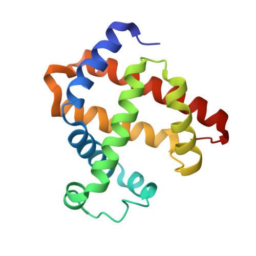
6E03

sperm whale myoglobin nitrosoethane adduct


Experimental Data Snapshot

  • Method: X-RAY DIFFRACTION
  • Resolution: 1.76 Å
  • R-Value Free: 0.191 
  • R-Value Work: 0.160 
  • R-Value Observed: 0.161 

wwPDB Validation   3D Report Full Report


Ligand Structure Quality Assessment 


This is version 1.2 of the entry. See complete history


Literature

Insights into Nitrosoalkane Binding to Myoglobin Provided by Crystallography of Wild-Type and Distal Pocket Mutant Derivatives.

Herrera, V.E.Charles, T.P.Scott, T.G.Prather, K.Y.Nguyen, N.T.Sohl, C.D.Thomas, L.M.Richter-Addo, G.B.

(2023) Biochemistry 62: 1406-1419

  • DOI: https://doi.org/10.1021/acs.biochem.2c00725
  • Primary Citation of Related Structures:  
    6E02, 6E03, 6E04, 8F9H, 8F9I, 8F9J, 8F9N

  • PubMed Abstract: 

    Nitrosoalkanes (R-N═O; R = alkyl) are biological intermediates that form from the oxidative metabolism of various amine (RNH 2 ) drugs or from the reduction of nitroorganics (RNO 2 ). RNO compounds bind to and inhibit various heme proteins. However, structural information on the resulting Fe-RNO moieties remains limited. We report the preparation of ferrous wild-type and H64A sw Mb II -RNO derivatives (λ max 424 nm; R = Me, Et, Pr, i Pr) from the reactions of Mb III -H 2 O with dithionite and nitroalkanes. The apparent extent of formation of the wt Mb derivatives followed the order MeNO > EtNO > PrNO > i PrNO, whereas the order was the opposite for the H64A derivatives. Ferricyanide oxidation of the Mb II -RNO derivatives resulted in the formation of the ferric Mb III -H 2 O precursors with loss of the RNO ligands. X-ray crystal structures of the wt Mb II -RNO derivatives at 1.76-2.0 Å resoln. revealed N -binding of RNO to Fe and the presence of H-bonding interactions between the nitroso O-atoms and distal pocket His64. The nitroso O-atoms pointed in the general direction of the protein exterior, and the hydrophobic R groups pointed toward the protein interior. X-ray crystal structures for the H64A mutant derivatives were determined at 1.74-1.80 Å resoln. An analysis of the distal pocket amino acid surface landscape provided an explanation for the differences in ligand orientations adopted by the EtNO and PrNO ligands in their wt and H64A structures. Our results provide a good baseline for the structural analysis of RNO binding to heme proteins possessing small distal pockets.


  • Organizational Affiliation

    Department of Chemistry and Physics, Ivory V. Nelson Science Center, Lincoln University, Lincoln University, Pennsylvania 19352, United States.


Macromolecules
Find similar proteins by:  (by identity cutoff)  |  3D Structure
Entity ID: 1
MoleculeChains Sequence LengthOrganismDetailsImage
Myoglobin154Physeter macrocephalusMutation(s): 0 
Gene Names: MB
EC: 1.11.1 (UniProt), 1.7 (UniProt)
UniProt
Find proteins for P02185 (Physeter macrocephalus)
Explore P02185 
Go to UniProtKB:  P02185
Entity Groups  
Sequence Clusters30% Identity50% Identity70% Identity90% Identity95% Identity100% Identity
UniProt GroupP02185
Sequence Annotations
Expand
  • Reference Sequence
Small Molecules
Ligands 4 Unique
IDChains Name / Formula / InChI Key2D Diagram3D Interactions
HEM
Query on HEM

Download Ideal Coordinates CCD File 
B [auth A]PROTOPORPHYRIN IX CONTAINING FE
C34 H32 Fe N4 O4
KABFMIBPWCXCRK-RGGAHWMASA-L
SO4
Query on SO4

Download Ideal Coordinates CCD File 
C [auth A],
D [auth A],
E [auth A],
F [auth A],
G [auth A]
SULFATE ION
O4 S
QAOWNCQODCNURD-UHFFFAOYSA-L
NOE
Query on NOE

Download Ideal Coordinates CCD File 
H [auth A]NITROSOETHANE
C2 H5 N O
IFYCAQIXDKZDTB-UHFFFAOYSA-N
CL
Query on CL

Download Ideal Coordinates CCD File 
I [auth A]CHLORIDE ION
Cl
VEXZGXHMUGYJMC-UHFFFAOYSA-M
Experimental Data & Validation

Experimental Data

  • Method: X-RAY DIFFRACTION
  • Resolution: 1.76 Å
  • R-Value Free: 0.191 
  • R-Value Work: 0.160 
  • R-Value Observed: 0.161 
  • Space Group: P 6
Unit Cell:
Length ( Å )Angle ( ˚ )
a = 90.272α = 90
b = 90.272β = 90
c = 45.316γ = 120
Software Package:
Software NamePurpose
REFMACrefinement
HKL-3000data reduction
HKL-3000data scaling
HKL-3000phasing

Structure Validation

View Full Validation Report


Ligand Structure Quality Assessment 


Entry History & Funding Information

Deposition Data


Funding OrganizationLocationGrant Number
National Science Foundation (NSF, United States)United States--

Revision History  (Full details and data files)

  • Version 1.0: 2019-07-17
    Type: Initial release
  • Version 1.1: 2019-11-27
    Changes: Author supporting evidence
  • Version 1.2: 2023-11-08
    Changes: Data collection, Database references