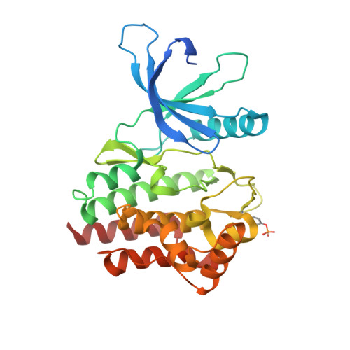
6VNL

JAK2 JH1 in complex with SG3-179


Experimental Data Snapshot

  • Method: X-RAY DIFFRACTION
  • Resolution: 2.40 Å
  • R-Value Free: 0.257 
  • R-Value Work: 0.217 
  • R-Value Observed: 0.218 

Starting Model: experimental
View more details

wwPDB Validation   3D Report Full Report


Ligand Structure Quality Assessment 


This is version 1.5 of the entry. See complete history


Literature

Structural Insights into JAK2 Inhibition by Ruxolitinib, Fedratinib, and Derivatives Thereof.

Davis, R.R.Li, B.Yun, S.Y.Chan, A.Nareddy, P.Gunawan, S.Ayaz, M.Lawrence, H.R.Reuther, G.W.Lawrence, N.J.Schonbrunn, E.

(2021) J Med Chem 64: 2228-2241

  • DOI: https://doi.org/10.1021/acs.jmedchem.0c01952
  • Primary Citation of Related Structures:  
    6VGL, 6VN8, 6VNB, 6VNC, 6VNE, 6VNF, 6VNG, 6VNH, 6VNI, 6VNJ, 6VNK, 6VNL, 6VNM, 6VS3, 6VSN

  • PubMed Abstract: 

    The discovery that aberrant activity of Janus kinase 2 (JAK2) is a driver of myeloproliferative neoplasms (MPNs) has led to significant efforts to develop small molecule inhibitors for this patient population. Ruxolitinib and fedratinib have been approved for use in MPN patients, while baricitinib, an achiral analogue of ruxolitinib, has been approved for rheumatoid arthritis. However, structural information on the interaction of these therapeutics with JAK2 remains unknown. Here, we describe a new methodology for the large-scale production of JAK2 from mammalian cells, which enabled us to determine the first crystal structures of JAK2 bound to these drugs and derivatives thereof. Along with biochemical and cellular data, the results provide a comprehensive view of the shape complementarity required for chiral and achiral inhibitors to achieve highest activity, which may facilitate the development of more effective JAK2 inhibitors as therapeutics.


  • Organizational Affiliation

    Drug Discovery DepartmentMoffitt Cancer Center, Tampa, Florida 33612, United States.


Macromolecules
Find similar proteins by:  (by identity cutoff)  |  3D Structure
Entity ID: 1
MoleculeChains Sequence LengthOrganismDetailsImage
Tyrosine-protein kinase JAK2A [auth D],
B,
C,
D [auth A]
308Homo sapiensMutation(s): 0 
Gene Names: JAK2
EC: 2.7.10.2
UniProt & NIH Common Fund Data Resources
Find proteins for O60674 (Homo sapiens)
Explore O60674 
Go to UniProtKB:  O60674
PHAROS:  O60674
GTEx:  ENSG00000096968 
Entity Groups  
Sequence Clusters30% Identity50% Identity70% Identity90% Identity95% Identity100% Identity
UniProt GroupO60674
Sequence Annotations
Expand
  • Reference Sequence
Small Molecules
Modified Residues  1 Unique
IDChains TypeFormula2D DiagramParent
PTR
Query on PTR
A [auth D],
B,
C,
D [auth A]
L-PEPTIDE LINKINGC9 H12 N O6 PTYR
Binding Affinity Annotations 
IDSourceBinding Affinity
5W2 BindingDB:  6VNL IC50: 11 (nM) from 1 assay(s)
Experimental Data & Validation

Experimental Data

  • Method: X-RAY DIFFRACTION
  • Resolution: 2.40 Å
  • R-Value Free: 0.257 
  • R-Value Work: 0.217 
  • R-Value Observed: 0.218 
  • Space Group: P 1 21 1
Unit Cell:
Length ( Å )Angle ( ˚ )
a = 111.76α = 90
b = 70.755β = 90.43
c = 112.861γ = 90
Software Package:
Software NamePurpose
PHENIXrefinement
PDB_EXTRACTdata extraction
XDSdata reduction
XDSdata scaling
PHASERphasing

Structure Validation

View Full Validation Report


Ligand Structure Quality Assessment 


Entry History & Funding Information

Deposition Data


Funding OrganizationLocationGrant Number
Leukemia & Lymphoma SocietyUnited States8012-18

Revision History  (Full details and data files)

  • Version 1.0: 2021-02-17
    Type: Initial release
  • Version 1.1: 2021-02-24
    Changes: Database references
  • Version 1.2: 2021-03-10
    Changes: Database references
  • Version 1.3: 2023-10-11
    Changes: Data collection, Database references, Refinement description
  • Version 1.4: 2023-11-15
    Changes: Data collection
  • Version 1.5: 2024-10-23
    Changes: Structure summary