6FUK

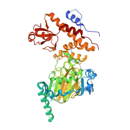
Crystal structure of UTX complexed with 5-carboxy-8-hydroxyquinoline


Experimental Data Snapshot

  • Method: X-RAY DIFFRACTION
  • Resolution: 2.00 Å
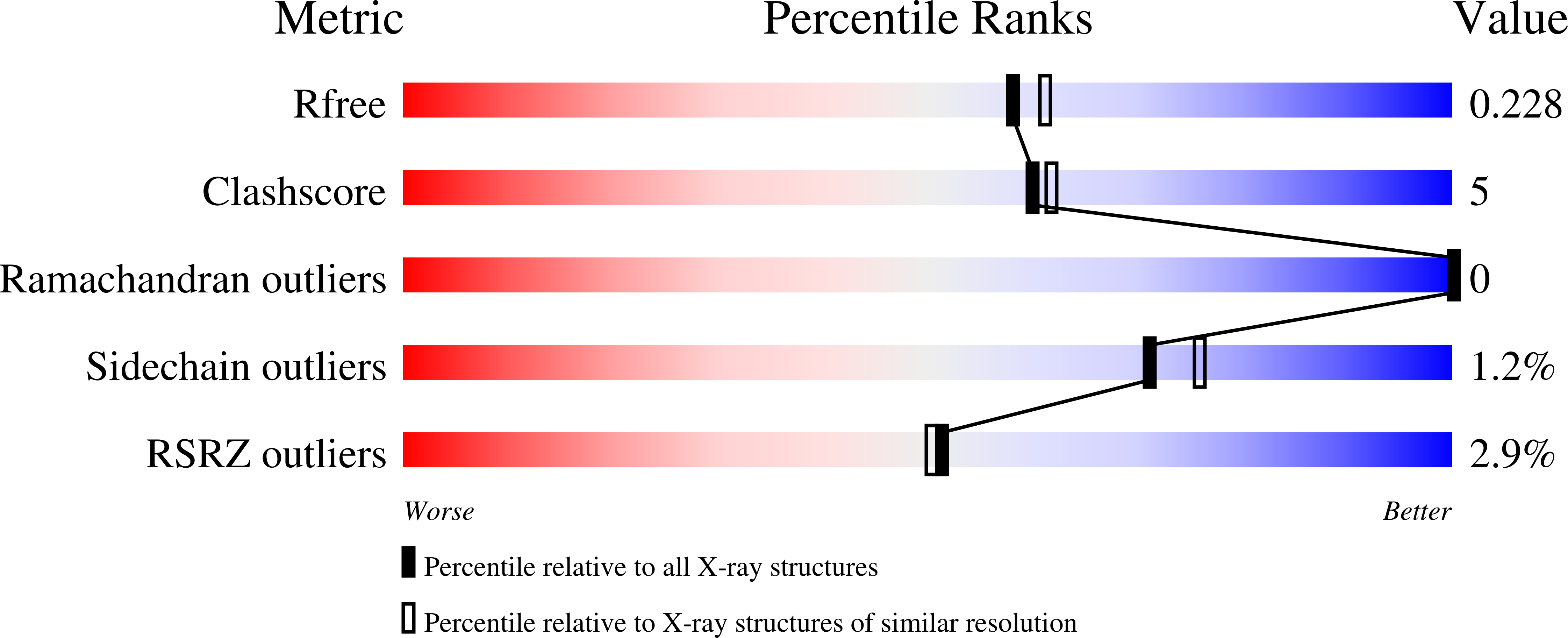
  • R-Value Free: 0.228 
  • R-Value Work: 0.186 
  • R-Value Observed: 0.188 

Starting Model: experimental
View more details

wwPDB Validation   3D Report Full Report


Ligand Structure Quality Assessment 


This is version 1.2 of the entry. See complete history


Literature

In Silico Identification of JMJD3 Demethylase Inhibitors.

Esposito, C.Wiedmer, L.Caflisch, A.

(2018) J Chem Inf Model 58: 2151-2163

  • DOI: https://doi.org/10.1021/acs.jcim.8b00539
  • Primary Citation of Related Structures:  
    6FUK, 6FUL, 6G8F

  • PubMed Abstract: 

    In the search for new demethylase inhibitors, we have developed a multistep protocol for in silico screening. Millions of poses generated by high-throughput docking or a 3D-pharmacophore search are first minimized by a classical force field and then filtered by semiempirical quantum mechanical calculations of the interaction energy with a selected set of functional groups in the binding site. The final ranking includes solvation effects which are evaluated in the continuum dielectric approximation (finite-difference Poisson equation). Application of the multistep protocol to JMJD3 jumonji demethylase has resulted in a dozen low-micromolar inhibitors belonging to five different chemical classes. We have solved the crystal structure of JMJD3 inhibitor 8 in the complex with UTX (a demethylase in the same subfamily as JMJD3) which validates the predicted binding mode. Compound 8 is a promising candidate for future optimization as it has a favorable ligand efficiency of 0.32 kcal/mol per nonhydrogen atom.


  • Organizational Affiliation

    Department of Biochemistry , University of Zurich , Winterthurerstrasse 190 , CH-8057 Zurich , Switzerland.


Macromolecules
Find similar proteins by:  (by identity cutoff)  |  3D Structure
Entity ID: 1
MoleculeChains Sequence LengthOrganismDetailsImage
Lysine-specific demethylase 6A531Homo sapiensMutation(s): 0 
Gene Names: KDM6AUTX
EC: 1.14.11 (PDB Primary Data), 1.14.11.68 (UniProt)
UniProt & NIH Common Fund Data Resources
Find proteins for O15550 (Homo sapiens)
Explore O15550 
Go to UniProtKB:  O15550
PHAROS:  O15550
GTEx:  ENSG00000147050 
Entity Groups  
Sequence Clusters30% Identity50% Identity70% Identity90% Identity95% Identity100% Identity
UniProt GroupO15550
Sequence Annotations
Expand
  • Reference Sequence
Small Molecules
Ligands 6 Unique
IDChains Name / Formula / InChI Key2D Diagram3D Interactions
8XQ
Query on 8XQ

Download Ideal Coordinates CCD File 
C [auth A]8-hydroxyquinoline-5-carboxylic acid
C10 H7 N O3
JGRPKOGHYBAVMW-UHFFFAOYSA-N
PG0
Query on PG0

Download Ideal Coordinates CCD File 
I [auth A]2-(2-METHOXYETHOXY)ETHANOL
C5 H12 O3
SBASXUCJHJRPEV-UHFFFAOYSA-N
PEG
Query on PEG

Download Ideal Coordinates CCD File 
G [auth A],
H [auth A]
DI(HYDROXYETHYL)ETHER
C4 H10 O3
MTHSVFCYNBDYFN-UHFFFAOYSA-N
SO4
Query on SO4

Download Ideal Coordinates CCD File 
E [auth A],
F [auth A]
SULFATE ION
O4 S
QAOWNCQODCNURD-UHFFFAOYSA-L
ZN
Query on ZN

Download Ideal Coordinates CCD File 
D [auth A]ZINC ION
Zn
PTFCDOFLOPIGGS-UHFFFAOYSA-N
MN
Query on MN

Download Ideal Coordinates CCD File 
B [auth A]MANGANESE (II) ION
Mn
WAEMQWOKJMHJLA-UHFFFAOYSA-N
Binding Affinity Annotations 
IDSourceBinding Affinity
8XQ BindingDB:  6FUK IC50: 200 (nM) from 1 assay(s)
Experimental Data & Validation

Experimental Data

  • Method: X-RAY DIFFRACTION
  • Resolution: 2.00 Å
  • R-Value Free: 0.228 
  • R-Value Work: 0.186 
  • R-Value Observed: 0.188 
  • Space Group: P 21 21 21
Unit Cell:
Length ( Å )Angle ( ˚ )
a = 80.417α = 90
b = 83.261β = 90
c = 92.898γ = 90
Software Package:
Software NamePurpose
PHENIXrefinement
XDSdata reduction
Cootmodel building
PHENIXphasing

Structure Validation

View Full Validation Report


Ligand Structure Quality Assessment 


Entry History 

Deposition Data

Revision History  (Full details and data files)

  • Version 1.0: 2018-10-10
    Type: Initial release
  • Version 1.1: 2018-10-31
    Changes: Data collection, Database references, Structure summary
  • Version 1.2: 2024-01-17
    Changes: Data collection, Database references, Derived calculations, Refinement description