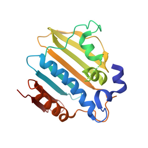
5Z9L

Bacterial GyrB ATPase domain in complex with a chemical fragment


Experimental Data Snapshot

  • Method: X-RAY DIFFRACTION
  • Resolution: 1.60 Å
  • R-Value Free: 0.242 
  • R-Value Work: 0.228 
  • R-Value Observed: 0.229 

Starting Model: experimental
View more details

wwPDB Validation   3D Report Full Report


Ligand Structure Quality Assessment 


This is version 1.1 of the entry. See complete history


Literature

Identification of an auxiliary druggable pocket in the DNA gyrase ATPase domain using fragment probes

Huang, X.Guo, J.Liu, Q.Gu, Q.Xu, J.Zhou, H.

(2018) Medchemcomm 9: 1619-1629

  • DOI: https://doi.org/10.1039/c8md00148k
  • Primary Citation of Related Structures:  
    5Z4H, 5Z4O, 5Z9B, 5Z9E, 5Z9F, 5Z9L, 5Z9M, 5Z9N, 5Z9P, 5Z9Q

  • PubMed Abstract: 

    Discovery of new drug binding sites on well-established targets is of great interest as it facilitates the design of new mechanistic inhibitors to overcome the acquired drug resistance. Small chemical fragments can easily enter and bind to the cavities on the protein surface. Thus, they can be used to probe new druggable pockets in proteins. DNA gyrase plays indispensable roles in DNA replication, and both its GyrA and GyrB subunits are clinically validated antibacterial targets. New mechanistic GyrB inhibitors are urgently desired since the withdrawal of novobiocin from the market by the FDA due to its reduced efficiency and other reasons. Here, a fragment library was screened against the E. coli GyrB ATPase domain by combining affinity- and bioactivity-based approaches. The following X-ray crystallographic efforts were made to determine the cocrystal structures of GyrB with ten fragment hits, and three different binding modes were disclosed. Fortunately, a hydrophobic pocket which is previously unknown was identified by two fragments. Fragments that bind to this pocket were shown to inhibit the ATPase activity as well as the DNA topological transition activity of DNA gyrase in vitro . A set of fragment analogs were screened to explore the binding capacity of this pocket and identify the better starting fragments for lead development. Phylogenetic analysis revealed that this pocket is conserved in most Gram-negative and also many Gram-positive human pathogenic bacteria, implying a broad-spectrum antibacterial potential and a lower risk of mutation. Thus, the novel druggable pocket and the starting fragments provide a novel basis for designing new GyrB-targeting therapeutics.


  • Organizational Affiliation

    Research Center for Drug Discovery , School of Pharmaceutical Sciences , Sun Yat-Sen University , Guangzhou 510006 , China . Email: [email protected].


Macromolecules
Find similar proteins by:  (by identity cutoff)  |  3D Structure
Entity ID: 1
MoleculeChains Sequence LengthOrganismDetailsImage
DNA gyrase subunit B
A, B
207Escherichia coli K-12Mutation(s): 0 
Gene Names: gyrBacrBcouhimBhisUnalCparApcbAb3699JW5625
EC: 5.99.1.3 (PDB Primary Data), 5.6.2.2 (UniProt)
UniProt
Find proteins for P0AES6 (Escherichia coli (strain K12))
Explore P0AES6 
Go to UniProtKB:  P0AES6
Entity Groups  
Sequence Clusters30% Identity50% Identity70% Identity90% Identity95% Identity100% Identity
UniProt GroupP0AES6
Sequence Annotations
Expand
  • Reference Sequence
Small Molecules
Ligands 3 Unique
IDChains Name / Formula / InChI Key2D Diagram3D Interactions
8H8 (Subject of Investigation/LOI)
Query on 8H8

Download Ideal Coordinates CCD File 
E [auth A],
H [auth B]
2-fluoro-4-hydroxybenzonitrile
C7 H4 F N O
REIVHYDACHXPNH-UHFFFAOYSA-N
AX7
Query on AX7

Download Ideal Coordinates CCD File 
I [auth B]1H-benzimidazol-2-amine
C7 H7 N3
JWYUFVNJZUSCSM-UHFFFAOYSA-N
PO4
Query on PO4

Download Ideal Coordinates CCD File 
C [auth A],
D [auth A],
F [auth B],
G [auth B]
PHOSPHATE ION
O4 P
NBIIXXVUZAFLBC-UHFFFAOYSA-K
Experimental Data & Validation

Experimental Data

  • Method: X-RAY DIFFRACTION
  • Resolution: 1.60 Å
  • R-Value Free: 0.242 
  • R-Value Work: 0.228 
  • R-Value Observed: 0.229 
  • Space Group: P 21 21 21
Unit Cell:
Length ( Å )Angle ( ˚ )
a = 61.07α = 90
b = 69.065β = 90
c = 102.121γ = 90
Software Package:
Software NamePurpose
REFMACrefinement
XDSdata reduction
SCALAdata scaling
MOLREPphasing

Structure Validation

View Full Validation Report


Ligand Structure Quality Assessment 


Entry History & Funding Information

Deposition Data


Funding OrganizationLocationGrant Number
National Natural Science Foundation of ChinaChina81773636

Revision History  (Full details and data files)

  • Version 1.0: 2018-12-12
    Type: Initial release
  • Version 1.1: 2023-11-22
    Changes: Data collection, Database references, Refinement description