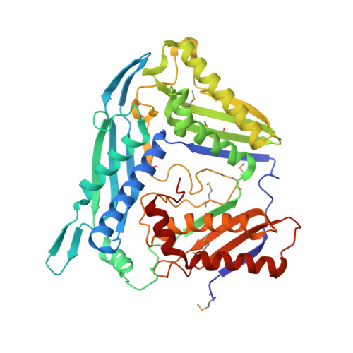
4L2Z

Crystal structure of S-Adenosylmethionine synthetase from Sulfolobus solfataricus complexed with SAE and PPi


Experimental Data Snapshot

  • Method: X-RAY DIFFRACTION
  • Resolution: 2.49 Å
  • R-Value Free: 0.184 
  • R-Value Work: 0.160 
  • R-Value Observed: 0.161 

Starting Model: experimental
View more details

wwPDB Validation   3D Report Full Report


Ligand Structure Quality Assessment 


This is version 1.6 of the entry. See complete history


Literature

Understanding molecular recognition of promiscuity of thermophilic methionine adenosyltransferase sMAT from Sulfolobus solfataricus.

Wang, F.Singh, S.Zhang, J.Huber, T.D.Helmich, K.E.Sunkara, M.Hurley, K.A.Goff, R.D.Bingman, C.A.Morris, A.J.Thorson, J.S.Phillips, G.N.

(2014) FEBS J 281: 4224-4239

  • DOI: https://doi.org/10.1111/febs.12784
  • Primary Citation of Related Structures:  
    4HPV, 4K0B, 4L2Z, 4L7I

  • PubMed Abstract: 

    Methionine adenosyltransferase (MAT) is a family of enzymes that utilizes ATP and methionine to produce S-adenosylmethionine (AdoMet), the most crucial methyl donor in the biological methylation of biomolecules and bioactive natural products. Here, we report that the MAT from Sulfolobus solfataricus (sMAT), an enzyme from a poorly explored class of the MAT family, has the ability to produce a range of differentially alkylated AdoMet analogs in the presence of non-native methionine analogs and ATP. To investigate the molecular basis for AdoMet analog production, we have crystallized the sMAT in the AdoMet bound, S-adenosylethionine (AdoEth) bound and unbound forms. Notably, among these structures, the AdoEth bound form offers the first MAT structure containing a non-native product, and cumulatively these structures add new structural insight into the MAT family and allow for detailed active site comparison with its homologs in Escherichia coli and human. As a thermostable MAT structure from archaea, the structures herein also provide a basis for future engineering to potentially broaden AdoMet analog production as reagents for methyltransferase-catalyzed 'alkylrandomization' and/or the study of methylation in the context of biological processes. PDB IDs: 4HPV, 4L7I, 4K0B and 4L2Z. EC 2.5.1.6 STRUCTURED DIGITAL ABSTRACT: • sMAT and sMAT bind by x-ray crystallography (View interaction).


  • Organizational Affiliation

    Department of Biochemistry and Cell Biology, Rice University, Houston, TX, USA.


Macromolecules
Find similar proteins by:  (by identity cutoff)  |  3D Structure
Entity ID: 1
MoleculeChains Sequence LengthOrganismDetailsImage
S-adenosylmethionine synthase
A, B
407Saccharolobus solfataricus P2Mutation(s): 0 
Gene Names: matSSO0199
EC: 2.5.1.6
UniProt
Find proteins for Q980S9 (Saccharolobus solfataricus (strain ATCC 35092 / DSM 1617 / JCM 11322 / P2))
Explore Q980S9 
Go to UniProtKB:  Q980S9
Entity Groups  
Sequence Clusters30% Identity50% Identity70% Identity90% Identity95% Identity100% Identity
UniProt GroupQ980S9
Sequence Annotations
Expand
  • Reference Sequence
Small Molecules
Ligands 4 Unique
IDChains Name / Formula / InChI Key2D Diagram3D Interactions
S7M
Query on S7M

Download Ideal Coordinates CCD File 
D [auth B][(3S)-3-amino-3-carboxypropyl]{[(2S,3S,4R,5R)-5-(6-amino-9H-purin-9-yl)-3,4-dihydroxytetrahydrofuran-2-yl]methyl}ethyls ulfonium
C16 H25 N6 O5 S
UBQZUBPODLPCFG-XIFWJBDBSA-O
DPO
Query on DPO

Download Ideal Coordinates CCD File 
E [auth B]DIPHOSPHATE
O7 P2
XPPKVPWEQAFLFU-UHFFFAOYSA-J
PO4
Query on PO4

Download Ideal Coordinates CCD File 
F [auth B]PHOSPHATE ION
O4 P
NBIIXXVUZAFLBC-UHFFFAOYSA-K
MG
Query on MG

Download Ideal Coordinates CCD File 
C [auth A],
G [auth B]
MAGNESIUM ION
Mg
JLVVSXFLKOJNIY-UHFFFAOYSA-N
Modified Residues  1 Unique
IDChains TypeFormula2D DiagramParent
MSE
Query on MSE
A, B
L-PEPTIDE LINKINGC5 H11 N O2 SeMET
Experimental Data & Validation

Experimental Data

Unit Cell:
Length ( Å )Angle ( ˚ )
a = 151.597α = 90
b = 151.597β = 90
c = 226.125γ = 120
Software Package:
Software NamePurpose
DENZOdata reduction
SCALEPACKdata scaling
PHENIXrefinement
PDB_EXTRACTdata extraction
HKL-2000data collection
HKL-2000data reduction
HKL-2000data scaling
PHENIXphasing

Structure Validation

View Full Validation Report


Ligand Structure Quality Assessment 


Entry History 

Revision History  (Full details and data files)

  • Version 1.0: 2013-06-19
    Type: Initial release
  • Version 1.1: 2014-04-02
    Changes: Database references
  • Version 1.2: 2014-10-08
    Changes: Database references
  • Version 1.3: 2017-11-15
    Changes: Refinement description
  • Version 1.4: 2023-09-20
    Changes: Data collection, Database references, Derived calculations, Refinement description, Structure summary
  • Version 1.5: 2023-12-06
    Changes: Data collection
  • Version 1.6: 2024-11-27
    Changes: Structure summary