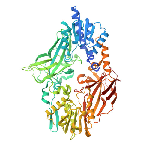
4C4A

Crystal structure of mouse protein arginine methyltransferase 7 in complex with SAH


Experimental Data Snapshot

  • Method: X-RAY DIFFRACTION
  • Resolution: 1.70 Å
  • R-Value Free: 0.190 
  • R-Value Work: 0.160 
  • R-Value Observed: 0.161 

Starting Model: experimental
View more details

wwPDB Validation   3D Report Full Report


Ligand Structure Quality Assessment 


This is version 1.4 of the entry. See complete history


Literature

Structural Insight Into Arginine Methylation by the Mouse Protein Arginine Methyltransferase 7: A Zinc Finger Freezes the Mimic of the Dimeric State Into a Single Active Site.

Cura, V.Troffer-Charlier, N.Wurtz, J.M.Bonnefond, L.Cavarelli, J.

(2014) Acta Crystallogr D Biol Crystallogr 70: 2401

  • DOI: https://doi.org/10.1107/S1399004714014278
  • Primary Citation of Related Structures:  
    4C4A

  • PubMed Abstract: 

    Protein arginine methyltransferase 7 (PRMT7) is a type III arginine methyltransferase which has been implicated in several biological processes such as transcriptional regulation, DNA damage repair, RNA splicing, cell differentiation and metastasis. PRMT7 is a unique but less characterized member of the family of PRMTs. The crystal structure of full-length PRMT7 from Mus musculus refined at 1.7 Å resolution is described. The PRMT7 structure is composed of two catalytic modules in tandem forming a pseudo-dimer and contains only one AdoHcy molecule bound to the N-terminal module. The high-resolution crystal structure presented here revealed several structural features showing that the second active site is frozen in an inactive state by a conserved zinc finger located at the junction between the two PRMT modules and by the collapse of two degenerated AdoMet-binding loops.


  • Organizational Affiliation

    Département de Biologie Structurale Intégrative, Institut de Génétique et de Biologie Moléculaire et Cellulaire (IGBMC), Université de Strasbourg, CNRS UMR7104, INSERM U596, 1 Rue Laurent Fries, F-67404 Illkirch, France.


Macromolecules
Find similar proteins by:  (by identity cutoff)  |  3D Structure
Entity ID: 1
MoleculeChains Sequence LengthOrganismDetailsImage
PROTEIN ARGININE N-METHYLTRANSFERASE 7692Mus musculusMutation(s): 0 
EC: 2.1.1 (PDB Primary Data), 2.1.1.125 (PDB Primary Data), 2.1.1.126 (PDB Primary Data), 2.1.1.321 (UniProt)
UniProt & NIH Common Fund Data Resources
Find proteins for Q922X9 (Mus musculus)
Explore Q922X9 
Go to UniProtKB:  Q922X9
IMPC:  MGI:2384879
Entity Groups  
Sequence Clusters30% Identity50% Identity70% Identity90% Identity95% Identity100% Identity
UniProt GroupQ922X9
Sequence Annotations
Expand
  • Reference Sequence
Small Molecules
Binding Affinity Annotations 
IDSourceBinding Affinity
SAH BindingDB:  4C4A IC50: min: 110, max: 5.00e+4 (nM) from 2 assay(s)
Experimental Data & Validation

Experimental Data

  • Method: X-RAY DIFFRACTION
  • Resolution: 1.70 Å
  • R-Value Free: 0.190 
  • R-Value Work: 0.160 
  • R-Value Observed: 0.161 
  • Space Group: P 43 21 2
Unit Cell:
Length ( Å )Angle ( ˚ )
a = 97.322α = 90
b = 97.322β = 90
c = 168.754γ = 90
Software Package:
Software NamePurpose
PHENIXrefinement
XDSdata reduction
Aimlessdata scaling
PHASERphasing

Structure Validation

View Full Validation Report


Ligand Structure Quality Assessment 


Entry History 

Deposition Data

Revision History  (Full details and data files)

  • Version 1.0: 2014-07-02
    Type: Initial release
  • Version 1.1: 2014-09-10
    Changes: Database references, Structure summary
  • Version 1.2: 2014-09-24
    Changes: Database references
  • Version 1.3: 2017-08-23
    Changes: Data collection
  • Version 1.4: 2024-05-01
    Changes: Data collection, Database references, Derived calculations, Other, Refinement description