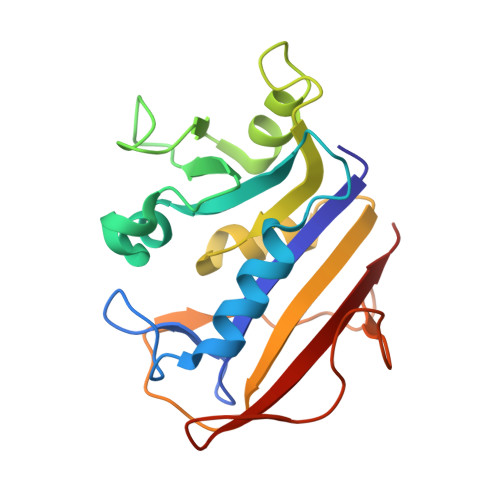
4KAK

Crystal structure of human dihydrofolate reductase complexed with NADPH and 6-ethyl-5-[(3S)-3-[3-methoxy-5-(pyridine-4-yl)phenyl]but-1-yn-1-yl]pyrimidine-2,4-diamine (UCP1006)


Experimental Data Snapshot

  • Method: X-RAY DIFFRACTION
  • Resolution: 1.80 Å
  • R-Value Free: 0.269 
  • R-Value Work: 0.223 
  • R-Value Observed: 0.225 

Starting Model: experimental
View more details

wwPDB Validation   3D Report Full Report


Ligand Structure Quality Assessment 


This is version 1.3 of the entry. See complete history


Literature

Elucidating features that drive the design of selective antifolates using crystal structures of human dihydrofolate reductase.

Lamb, K.M.G-Dayanandan, N.Wright, D.L.Anderson, A.C.

(2013) Biochemistry 52: 7318-7326

  • DOI: https://doi.org/10.1021/bi400852h
  • Primary Citation of Related Structures:  
    4KAK, 4KBN, 4KD7, 4KEB, 4KFJ

  • PubMed Abstract: 

    The pursuit of antimicrobial drugs that target dihydrofolate reductase (DHFR) exploits differences in sequence and dynamics between the pathogenic and human enzymes. Here, we present five crystal structures of human DHFR bound to a new class of antimicrobial agents, the propargyl-linked antifolates (PLAs), with a range of potency (IC50 values of 0.045-1.07 μM) for human DHFR. These structures reveal that interactions between the ligands and Asn 64, Phe 31, and Phe 34 are important for increased affinity for human DHFR and that loop residues 58-64 undergo ligand-induced conformational changes. The utility of these structural studies was demonstrated through the design of three new ligands that reduce the number of contacts with Asn 64, Phe 31, and Phe 34. Synthesis and evaluation show that one of the designed inhibitors exhibits the lowest affinity for human DHFR of any of the PLAs (2.64 μM). Comparisons of structures of human and Staphylococcus aureus DHFR bound to the same PLA reveal a conformational change in the ligand that enhances interactions with residues Phe 92 (Val 115 in huDHFR) and Ile 50 (Ile 60 in huDHFR) in S. aureus DHFR, yielding selectivity. Likewise, comparisons of human and Candida glabrata DHFR bound to the same ligand show that hydrophobic interactions with residues Ile 121 and Phe 66 (Val 115 and Asn 64 in human DHFR) yield selective inhibitors. The identification of residue substitutions that are important for selectivity and the observation of active site flexibility will help guide antimicrobial antifolate development for the inhibition of pathogenic species.


  • Organizational Affiliation

    Department of Pharmaceutical Sciences, University of Connecticut , 69 North Eagleville Road, Storrs, Connecticut 06269, United States.


Macromolecules
Find similar proteins by:  (by identity cutoff)  |  3D Structure
Entity ID: 1
MoleculeChains Sequence LengthOrganismDetailsImage
Dihydrofolate reductase
A, B
186Homo sapiensMutation(s): 0 
Gene Names: DHFRhuDHFR
EC: 1.5.1.3
UniProt & NIH Common Fund Data Resources
Find proteins for P00374 (Homo sapiens)
Explore P00374 
Go to UniProtKB:  P00374
PHAROS:  P00374
GTEx:  ENSG00000228716 
Entity Groups  
Sequence Clusters30% Identity50% Identity70% Identity90% Identity95% Identity100% Identity
UniProt GroupP00374
Sequence Annotations
Expand
  • Reference Sequence
Small Molecules
Ligands 4 Unique
IDChains Name / Formula / InChI Key2D Diagram3D Interactions
NDP
Query on NDP

Download Ideal Coordinates CCD File 
C [auth A],
I [auth B]
NADPH DIHYDRO-NICOTINAMIDE-ADENINE-DINUCLEOTIDE PHOSPHATE
C21 H30 N7 O17 P3
ACFIXJIJDZMPPO-NNYOXOHSSA-N
06U
Query on 06U

Download Ideal Coordinates CCD File 
D [auth A],
J [auth B]
6-ethyl-5-{(3R)-3-[3-methoxy-5-(pyridin-4-yl)phenyl]but-1-yn-1-yl}pyrimidine-2,4-diamine
C22 H23 N5 O
KEPLBUUTAQCZOE-AWEZNQCLSA-N
EOH
Query on EOH

Download Ideal Coordinates CCD File 
E [auth A],
F [auth A],
G [auth A],
K [auth B],
L [auth B]
ETHANOL
C2 H6 O
LFQSCWFLJHTTHZ-UHFFFAOYSA-N
CA
Query on CA

Download Ideal Coordinates CCD File 
H [auth A],
M [auth B],
N [auth B],
O [auth B]
CALCIUM ION
Ca
BHPQYMZQTOCNFJ-UHFFFAOYSA-N
Binding Affinity Annotations 
IDSourceBinding Affinity
06U BindingDB:  4KAK IC50: 1300 (nM) from 1 assay(s)
Experimental Data & Validation

Experimental Data

  • Method: X-RAY DIFFRACTION
  • Resolution: 1.80 Å
  • R-Value Free: 0.269 
  • R-Value Work: 0.223 
  • R-Value Observed: 0.225 
  • Space Group: C 2 2 21
Unit Cell:
Length ( Å )Angle ( ˚ )
a = 88.077α = 90
b = 93.634β = 90
c = 96.039γ = 90
Software Package:
Software NamePurpose
StructureStudiodata collection
PHASERphasing
PHENIXrefinement

Structure Validation

View Full Validation Report


Ligand Structure Quality Assessment 


Entry History 

Deposition Data

Revision History  (Full details and data files)

  • Version 1.0: 2013-10-09
    Type: Initial release
  • Version 1.1: 2013-10-30
    Changes: Database references
  • Version 1.2: 2013-11-20
    Changes: Non-polymer description
  • Version 1.3: 2023-09-20
    Changes: Data collection, Database references, Derived calculations, Refinement description