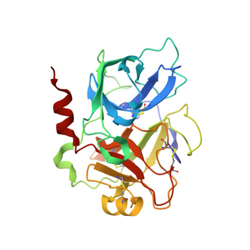
4CRF

Creating novel F1 inhibitors through fragment based lead generation and structure aided drug design


Experimental Data Snapshot

  • Method: X-RAY DIFFRACTION
  • Resolution: 2.30 Å
  • R-Value Free: 0.244 
  • R-Value Work: 0.181 
  • R-Value Observed: 0.184 

wwPDB Validation   3D Report Full Report


Ligand Structure Quality Assessment 


This is version 2.1 of the entry. See complete history


Literature

Creating Novel Activated Factor Xi Inhibitors Through Fragment Based Lead Generation and Structure Aided Drug Design.

Fjellstrom, O.Akkaya, S.Beisel, H.Eriksson, P.Erixon, K.Gustafsson, D.Jurva, U.Kang, D.Karis, D.Knecht, W.Nerme, V.Nilsson, I.Olsson, T.Redzic, A.Roth, R.Sandmark, J.Tigerstrom, A.Oster, L.

(2015) PLoS One 10: 13705

  • DOI: https://doi.org/10.1371/journal.pone.0113705
  • Primary Citation of Related Structures:  
    4CR5, 4CR9, 4CRA, 4CRB, 4CRC, 4CRD, 4CRE, 4CRF, 4CRG

  • PubMed Abstract: 

    Activated factor XI (FXIa) inhibitors are anticipated to combine anticoagulant and profibrinolytic effects with a low bleeding risk. This motivated a structure aided fragment based lead generation campaign to create novel FXIa inhibitor leads. A virtual screen, based on docking experiments, was performed to generate a FXIa targeted fragment library for an NMR screen that resulted in the identification of fragments binding in the FXIa S1 binding pocket. The neutral 6-chloro-3,4-dihydro-1H-quinolin-2-one and the weakly basic quinolin-2-amine structures are novel FXIa P1 fragments. The expansion of these fragments towards the FXIa prime side binding sites was aided by solving the X-ray structures of reported FXIa inhibitors that we found to bind in the S1-S1'-S2' FXIa binding pockets. Combining the X-ray structure information from the identified S1 binding 6-chloro-3,4-dihydro-1H-quinolin-2-one fragment and the S1-S1'-S2' binding reference compounds enabled structure guided linking and expansion work to achieve one of the most potent and selective FXIa inhibitors reported to date, compound 13, with a FXIa IC50 of 1.0 nM. The hydrophilicity and large polar surface area of the potent S1-S1'-S2' binding FXIa inhibitors compromised permeability. Initial work to expand the 6-chloro-3,4-dihydro-1H-quinolin-2-one fragment towards the prime side to yield molecules with less hydrophilicity shows promise to afford potent, selective and orally bioavailable compounds.


  • Organizational Affiliation

    Medicinal Chemistry, Cardiovascular & Metabolic Diseases Innovative Medicines, AstraZeneca R&D, Mölndal, Sweden.


Macromolecules
Find similar proteins by:  (by identity cutoff)  |  3D Structure
Entity ID: 1
MoleculeChains Sequence LengthOrganismDetailsImage
Coagulation factor XIa light chain238Homo sapiensMutation(s): 0 
Gene Names: F11
EC: 3.4.21.27
UniProt & NIH Common Fund Data Resources
Find proteins for P03951 (Homo sapiens)
Explore P03951 
Go to UniProtKB:  P03951
PHAROS:  P03951
GTEx:  ENSG00000088926 
Entity Groups  
Sequence Clusters30% Identity50% Identity70% Identity90% Identity95% Identity100% Identity
UniProt GroupP03951
Sequence Annotations
Expand
  • Reference Sequence
Small Molecules
Ligands 4 Unique
IDChains Name / Formula / InChI Key2D Diagram3D Interactions
R9B
Query on R9B

Download Ideal Coordinates CCD File 
E [auth A]N-[(1S)-1-benzyl-2-[(6-chloro-2-oxo-1H-quinolin-4-yl)methylamino]-2-oxo-ethyl]-4-hydroxy-2-oxo-1H-quinoline-6-carbo
C29 H23 Cl N4 O5
DNDMLXSODUEUMP-DEOSSOPVSA-N
SO4
Query on SO4

Download Ideal Coordinates CCD File 
F [auth A],
G [auth A],
H [auth A],
I [auth A]
SULFATE ION
O4 S
QAOWNCQODCNURD-UHFFFAOYSA-L
GOL
Query on GOL

Download Ideal Coordinates CCD File 
B [auth A],
C [auth A],
D [auth A]
GLYCEROL
C3 H8 O3
PEDCQBHIVMGVHV-UHFFFAOYSA-N
CL
Query on CL

Download Ideal Coordinates CCD File 
J [auth A]CHLORIDE ION
Cl
VEXZGXHMUGYJMC-UHFFFAOYSA-M
Binding Affinity Annotations 
IDSourceBinding Affinity
R9B BindingDB:  4CRF Ki: 0.5 (nM) from 1 assay(s)
IC50: 1 (nM) from 1 assay(s)
Experimental Data & Validation

Experimental Data

  • Method: X-RAY DIFFRACTION
  • Resolution: 2.30 Å
  • R-Value Free: 0.244 
  • R-Value Work: 0.181 
  • R-Value Observed: 0.184 
  • Space Group: I 2 3
Unit Cell:
Length ( Å )Angle ( ˚ )
a = 120.99α = 90
b = 120.99β = 90
c = 120.99γ = 90
Software Package:
Software NamePurpose
REFMACrefinement
MOSFLMdata reduction
SCALAdata scaling
MOLREPphasing

Structure Validation

View Full Validation Report


Ligand Structure Quality Assessment 


Entry History 

Revision History  (Full details and data files)

  • Version 1.0: 2015-02-11
    Type: Initial release
  • Version 1.1: 2018-04-04
    Changes: Data collection
  • Version 1.2: 2019-03-06
    Changes: Data collection, Experimental preparation
  • Version 2.0: 2022-05-04
    Changes: Advisory, Atomic model, Data collection, Database references, Derived calculations, Other, Source and taxonomy, Structure summary
  • Version 2.1: 2024-11-06
    Changes: Data collection, Structure summary