3CA3

Crystal structure of Sambucus Nigra Agglutinin II (SNA-II)-tetragonal crystal form- complexed to N-Acetylgalactosamine


Experimental Data Snapshot

  • Method: X-RAY DIFFRACTION
  • Resolution: 1.55 Å
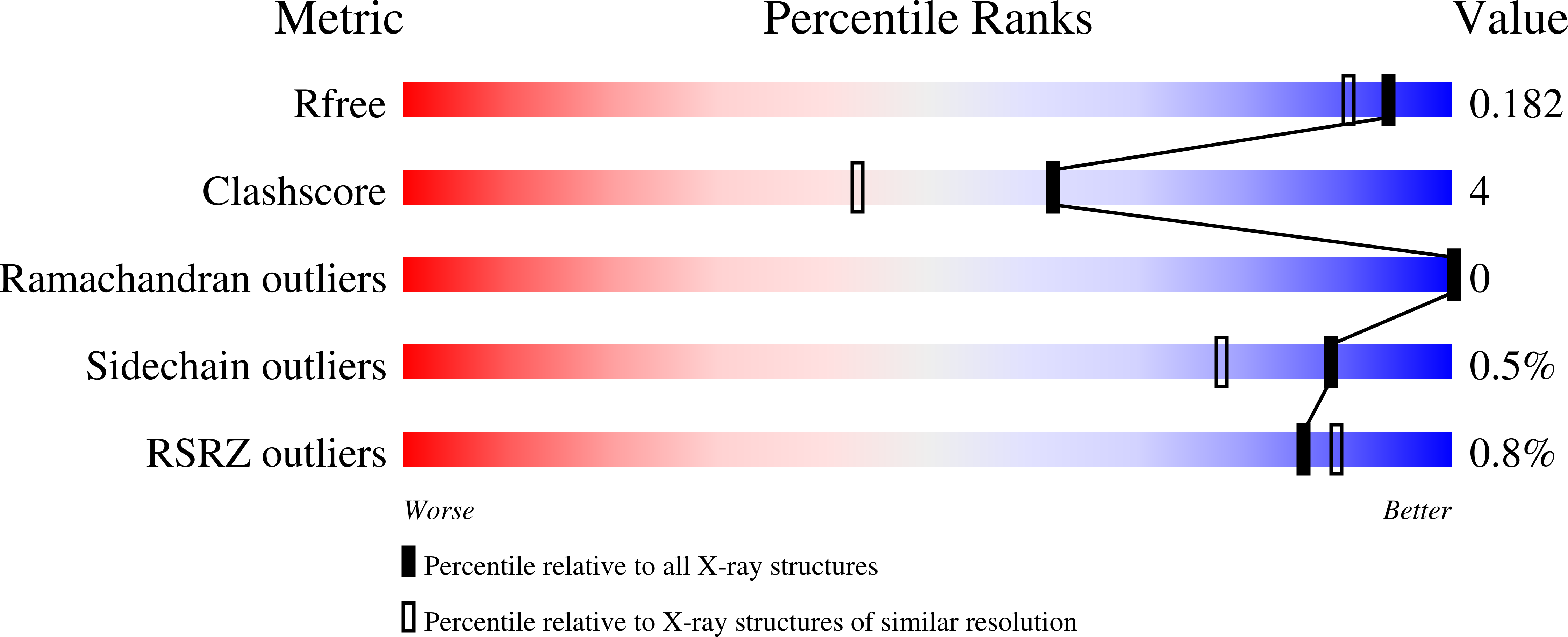
  • R-Value Free: 0.173 
  • R-Value Work: 0.122 
  • R-Value Observed: 0.125 

Starting Model: experimental
View more details

wwPDB Validation   3D Report Full Report


Ligand Structure Quality Assessment 


This is version 2.2 of the entry. See complete history


Literature

Structural basis for sugar recognition, including the Tn carcinoma antigen, by the lectin SNA-II from Sambucus nigra

Maveyraud, L.Niwa, H.Guillet, V.Svergun, D.I.Konarev, P.V.Palmer, R.A.Peumans, W.J.Rouge, P.Van Damme, E.J.Reynolds, C.D.Mourey, L.

(2009) Proteins 75: 89-103

  • DOI: https://doi.org/10.1002/prot.22222
  • Primary Citation of Related Structures:  
    3C9Z, 3CA0, 3CA1, 3CA3, 3CA4, 3CA5, 3CA6, 3CAH

  • PubMed Abstract: 

    Bark of elderberry (Sambucus nigra) contains a galactose (Gal)/N-acetylgalactosamine (GalNAc)-specific lectin (SNA-II) corresponding to slightly truncated B-chains of a genuine Type-II ribosome-inactivating protein (Type-II RIPs, SNA-V), found in the same species. The three-dimensional X-ray structure of SNA-II has been determined in two distinct crystal forms, hexagonal and tetragonal, at 1.90 A and 1.35 A, respectively. In both crystal forms, the SNA-II molecule folds into two linked beta-trefoil domains, with an overall conformation similar to that of the B-chains of ricin and other Type-II RIPs. Glycosylation is observed at four sites along the polypeptide chain, accounting for 14 saccharide units. The high-resolution structures of SNA-II in complex with Gal and five Gal-related saccharides (GalNAc, lactose, alpha1-methylgalactose, fucose, and the carcinoma-specific Tn antigen) were determined at 1.55 A resolution or better. Binding is observed in two saccharide-binding sites for most of the sugars: a conserved aspartate residue interacts simultaneously with the O3 and O4 atoms of saccharides. In one of the binding sites, additional interactions with the protein involve the O6 atom. Analytical gel filtration, small angle X-ray scattering studies and crystal packing analysis indicate that, although some oligomeric species are present, the monomeric species predominate in solution.


  • Organizational Affiliation

    Institut de Pharmacologie et de Biologie Structurale (IPBS), UMR 5089, Université Paul Sabatier Toulouse III/CNRS, Toulouse, France. [email protected]


Macromolecules
Find similar proteins by:  (by identity cutoff)  |  3D Structure
Entity ID: 1
MoleculeChains Sequence LengthOrganismDetailsImage
Agglutinin II258Sambucus nigraMutation(s): 0 
UniProt
Find proteins for P33183 (Sambucus nigra)
Explore P33183 
Go to UniProtKB:  P33183
Entity Groups  
Sequence Clusters30% Identity50% Identity70% Identity90% Identity95% Identity100% Identity
UniProt GroupP33183
Glycosylation
Glycosylation Sites: 4
Sequence Annotations
Expand
  • Reference Sequence
Oligosaccharides

Help

Entity ID: 2
MoleculeChains Length2D Diagram Glycosylation3D Interactions
alpha-L-fucopyranose-(1-3)-[2-acetamido-2-deoxy-beta-D-glucopyranose-(1-4)]2-acetamido-2-deoxy-beta-D-glucopyranose
B, C
3N-Glycosylation
Glycosylation Resources
GlyTouCan:  G11971MR
GlyCosmos:  G11971MR
GlyGen:  G11971MR
Entity ID: 3
MoleculeChains Length2D Diagram Glycosylation3D Interactions
2-acetamido-2-deoxy-beta-D-glucopyranose-(1-4)-2-acetamido-2-deoxy-beta-D-glucopyranose
D
2N-Glycosylation
Glycosylation Resources
GlyTouCan:  G42666HT
GlyCosmos:  G42666HT
GlyGen:  G42666HT
Small Molecules
Ligands 5 Unique
IDChains Name / Formula / InChI Key2D Diagram3D Interactions
A2G
Query on A2G

Download Ideal Coordinates CCD File 
F [auth A],
G [auth A]
2-acetamido-2-deoxy-alpha-D-galactopyranose
C8 H15 N O6
OVRNDRQMDRJTHS-CBQIKETKSA-N
NGA
Query on NGA

Download Ideal Coordinates CCD File 
H [auth A],
I [auth A]
2-acetamido-2-deoxy-beta-D-galactopyranose
C8 H15 N O6
OVRNDRQMDRJTHS-JAJWTYFOSA-N
NAG
Query on NAG

Download Ideal Coordinates CCD File 
E [auth A]2-acetamido-2-deoxy-beta-D-glucopyranose
C8 H15 N O6
OVRNDRQMDRJTHS-FMDGEEDCSA-N
SO4
Query on SO4

Download Ideal Coordinates CCD File 
J [auth A]
K [auth A]
L [auth A]
M [auth A]
N [auth A]
J [auth A],
K [auth A],
L [auth A],
M [auth A],
N [auth A],
O [auth A],
P [auth A]
SULFATE ION
O4 S
QAOWNCQODCNURD-UHFFFAOYSA-L
ACT
Query on ACT

Download Ideal Coordinates CCD File 
Q [auth A]ACETATE ION
C2 H3 O2
QTBSBXVTEAMEQO-UHFFFAOYSA-M
Experimental Data & Validation

Experimental Data

  • Method: X-RAY DIFFRACTION
  • Resolution: 1.55 Å
  • R-Value Free: 0.173 
  • R-Value Work: 0.122 
  • R-Value Observed: 0.125 
  • Space Group: I 41 2 2
Unit Cell:
Length ( Å )Angle ( ˚ )
a = 126.124α = 90
b = 126.124β = 90
c = 76.039γ = 90
Software Package:
Software NamePurpose
REFMACrefinement
DNAdata collection
MOSFLMdata reduction
SCALAdata scaling
REFMACphasing

Structure Validation

View Full Validation Report


Ligand Structure Quality Assessment 


Entry History 

Deposition Data

Revision History  (Full details and data files)

  • Version 1.0: 2008-11-25
    Type: Initial release
  • Version 1.1: 2011-07-13
    Changes: Derived calculations, Version format compliance
  • Version 2.0: 2020-07-29
    Type: Remediation
    Reason: Carbohydrate remediation
    Changes: Atomic model, Data collection, Derived calculations, Structure summary
  • Version 2.1: 2024-04-03
    Changes: Data collection, Database references, Refinement description, Structure summary
  • Version 2.2: 2024-11-20
    Changes: Structure summary